-
 Call Now ! +31 88 3886069

Our department offers an infrastructure that covers advanced in vitro and in vivo models, state-of-the-art analytical techniques, and cutting-edge imaging, molecular, and computational tools. Together, these platforms enable mechanistic, translational, and exposure-driven research for a wide range of biomedical and environmental health questions. The listed techniques are available and can be combined into tailored experimental pipelines, supporting both collaborative projects and service-oriented studies.
For further information, please contact secretariaat-phartox@maastrichtuniversity.nl, who will be happy to assist you and direct your inquiry to the appropriate experts.
Advanced Cell Culture & In Vitro Models
Various 2D cell culture models
Human primary cell cultures
3D human cell culture models
- Intestinal epithelial models
- Bronchial and alveolar epithelial models
Air–liquid interface (ALI) culture systems
Endothelial cell culture models
- Barrier integrity (TEER)
- Permeability assays
Exposure & Inhalation
Air-liquid interface exposure systems (static)
Nanoparticle generation for toxicity studies (spark ablation)
Particle characterization techniques
Genome Stability
Comet assay (single-cell gel electrophoresis)
- Standard and enzyme-modified versions
- High-throughput formats
- Applications in cells, whole blood, and tissues)
DNA repair capacity assays
- Base excision repair (BER)
- Nucleotide excision repair (NER)
Micronucleus assay (chromosomal damage)
yH2AX staining (DNA double-strand breaks)
DNA adduct analysis
- LC-MS–based approaches
- Gel electrophoresis–based methods)
Telomere length analysis
Mitochondrial DNA damage and copy number analysis
Molecular Biology & Cellular Signaling
Standard molecular biology assays
- PCR / qPCR
- Western blot
- ELISA
Intracellular signaling pathway analysis
- Inflammation (e.g. NF-kB, AP-1)
- Oxidative stress pathways
- Mitochondrial biogenesis and mitophagy
Autophagy and mitochondrial function analyses
Redox biology assays
- Antioxidant enzyme activity
- Oxidative stress read-outs
Cytochrome P450 enzyme activity assays (e.g. CYP1A1 / EROD)
AhR-CALUX reporter gene assay
Nutritional Biochemistry and Digestion & Effect-Based Analysis
INFOGEST static in vitro digestion models
-Simulation of oral, gastric, and intestinal phases
- Assessment of protein digestibility and peptide release
Metabolomics & Chemical Analysis
LC-HRMS–based metabolomics
HPLC with fluorescence detection
Volatile organic compound (VOC) analysis
- High-resolution GC-Orbitrap-MS
- Targeted and non-targeted workflows
Sorptive extraction techniques (HiSorb(tm))
Thermal desorption–GC-MS
Time-resolved VOC emission profiling
VOC analysis in complex matrices
- Biological samples
- Environmental samples
- Materials
Integrated sampling, enrichment, and accurate-mass identification
Calculation of protein quality indices (e.g., DIAAS-related metrics)
Analysis in food matrices, digesta, and biological samples
Spectrophotometric quantification assays for:
- Phytates
- Tannins
- Total phenolics
Assessment of nutrient–antinutrient interactions
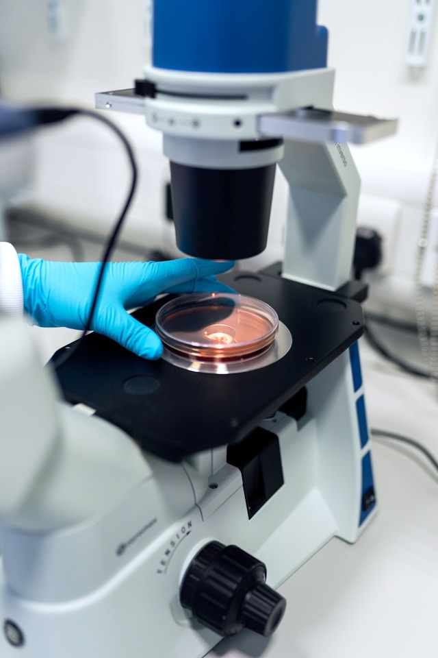
Imaging, Microscopy & Spectroscopy
Live-cell imaging platforms
Advanced imaging modalities
- Laser Speckle Contrast Imaging (cerebral blood flow)
In vivo Models & Physiology
Animal models of vascular cognitive impairment and dementia
Cardiovascular phenotyping
- Blood pressure monitoring
- Arterial stiffness and remodeling
Cerebral blood flow and neurovascular coupling assessment
Brain microvessel isolation and characterization
Vascular Function & Hemodynamics
Wire myography (arterial contractility and endothelial function)
Bioinformatics & Data Integration
Automated image analysis (2D & 3D)
Machine-learning–based image quantification
Bioinformatic pipelines for large (epi)genetic datasets
Supervised and unsupervised machine learning approaches for large data sets
Metabolomics-based machine learning analysis
Additionally, we offer techniques that are executed using our expertise while leveraging facilities elsewhere at Maastricht University. Although the experiments are carried out by our team, the resources belong to the wider university infrastructure, allowing access to specialized platforms that complement our in-house capabilities.
Exposure & Inhalation
Air-liquid interface exposure systems (continuous-flow)
Metabolomics & Chemical Analysis
Total amino acid determination
Imaging, Microscopy & Spectroscopy
Automated fluorescent microscopy scanning
Confocal and slide-scanning microscopy
Electron microscopy (EM)
Light-sheet microscopy of cleared tissues
Spectroscopic techniques
- Electron spin resonance (ESR)
In vivo Models & Physiology
Cognitive and behavioral testing
- Memory and learning paradigms
In vivo metabolic phenotyping
- Hyperinsulinemic–euglycemic clamp
Ultrasound-based microvascular imaging
Vascular Function & Hemodynamics
Pressure myography (resistance artery structure and function)
Pulse wave velocity measurements
Arterial wall thickness assessment
Contrast-enhanced ultrasound for microvascular perfusion
Bioinformatics & Data Integration
Transcriptomic analysis
- Bulk RNA-sequencing
- Primary cells, tissues, and microvessels